

Dra. Karen Fernandes de Oliveira

Existem diversas condições que cursam com queda de cabelo e que podem imitar a alopecia androgenética (AGA), representando um desafio diagnóstico e terapêutico para cirurgiões de transplante capilar. A alopecia androgenética, uma das causas mais comuns de queda de cabelo, caracteriza-se por um padrão hereditário em homens e mulheres e geralmente responde bem aos tratamentos clínicos e cirúrgico. No entanto, outras alopecias, tanto cicatriciais quanto não cicatriciais, podem se assemelhar a AGA, mas não respondem de forma tão eficaz ao transplante capilar. Algumas dessas condições podem não permitir crescimento capilar satisfatório após o transplante, gerando frustração para pacientes e médicos.
Entre as alopecias não cicatriciais, a alopecia areata (AA) é uma condição autoimune que pode ser confundida com AGA, especialmente quando ocorre na região do vértex. A AA difusa apresenta uma perda de cabelo mais ampla e uniforme podendo ser confundida com a AGA difusa em casos crônicos. O diagnóstico pode ser auxiliado por tricoscopia, que identifica características como pontos amarelos, pontos pretos e fios peládicos (em ponto de exclamação). (Imagem 1). Apesar de existirem tratamentos com esteroides tópicos ou sistêmicos e imunossupressores para o controle da alopecia, o transplante capilar em pacientes com AA é desaconselhado.
As alopecias cicatriciais, por sua vez, são especialmente difíceis de tratar com transplante devido à destruição dos folículos pilosos e à natureza inflamatória. Alopecias como a alopecia frontal fibrosante (AFF) em região frontal e líquen plano pilar (LPP) em região de vértex, por exemplo, podem simular uma AGA ao exame clínico, porém causam cicatrizes permanentes e exigem controle rigoroso da inflamação antes de se considerar o transplante. Na AFF, o recuo da linha frontal e a perda de sobrancelhas são sinais típicos, e são necessários tratamentos sistêmicos com anti andrógenos, anti-inflamatórios e por vezes até imunossupressores além de terapias tópicas para conter a progressão e assegurar estabilidade da doença por anos para avaliar a possibilidade de uma cirurgia de transplante. Ainda sim, é necessário conscientizar o paciente de que embora os resultados a curto prazo possam ser satisfatórios, a perda progressiva dos fios transplantados pode ocorrer ao longo do tempo: mais de 50 % dos fios transplantados podem ser perdidos em até 5 anos após a cirurgia nos pacientes com AFF.
A avaliação do couro cabeludo com tricoscopia e uma biópsia são recomendadas antes de um transplante capilar para distinguir entre AGA e suas condições imitadoras, especialmente nos casos de suspeita de alopecia cicatricial, nos quais o transplante não é a primeira escolha de tratamento. A tricoscopia é uma ferramenta valiosa que permite a visualização com aumento do couro cabeludo e dos fios de cabelo, ajudando a distinguir entre diferentes tipos de alopecia. Na alopecia androgenética há anisotricose de fios terminais, aumento de velos, fios miniaturizados e pontos amarelos (Imagem 2), enquanto no LPP e na AFF, pode-se observar eritema e descamação peripilar, pilli torti, pontos brancos fibróticos e eritema difuso ao exame tricoscópico. (Imagem 3).
Além disso, na avaliação histopatológica, observa-se diminuição da contagem folicular, ausência de glândulas sebáceas infiltrado linfocítico e fibrose perifolicular - informações dadas pelo dermatopatologista que diferenciam o LPP e a AFF da AGA.
Por fim, é essencial que os cirurgiões de transplante capilar estejam atentos aos sinais de inflamação do couro cabeludo, realizem a tricoscopia antes da cirurgia e, se necessário, a biópsia do couro cabeludo. A abordagem cautelosa e a comunicação clara com o paciente sobre as possibilidades e limitações do transplante capilar nesses casos são fundamentais para minimizar decepções e alinhar expectativas quanto à cirurgia de transplante capilar.
Imagem 1: alopecia areata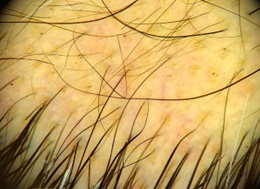
Imagem 2: alopecia androgenética masculina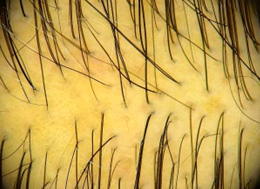
Imagem 3: líquen plano pilar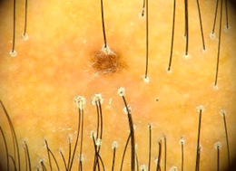
Referências bibliográficas sugeridas:
- KHUTSISHVILI, N.; RUDNICKA, L.; OVCHARENKO, Y.; et al. Trichoscopy - A valuable tool for identifying conditions mimicking androgenetic alopecia. International Journal of Dermatology, v. 63, n. 1, p. 23-31, 2024. DOI: 10.1111/ijd.16895.
- OLIVEIRA, K. F. Tricoscopia: o que o cirurgião de calvície precisa saber. In: MURICY, M. A.; MURICY, J. C. Métodos Avançados de Transplante Capilar FUE. 1. ed. Rio de Janeiro: Dilivros Editora, 2024. p. 39-48.
- MUBKI, T.; RUDNICKA, L.; OLSZEWSKA, M.; SHAPIRO, J. Evaluation and diagnosis of the hair loss patient: part II. Trichoscopic and laboratory evaluations. Journal of the American Academy of Dermatology, v. 71, n. 3, p. 431.e1-431.e11, 2014. DOI: 10.1016/j.jaad.2014.05.008.
- RUDNICKA, L.; RAKOWSKA, A.; KERZEJA, M.; OLSZEWSKA, M. Hair shafts in trichoscopy: clues for diagnosis of hair and scalp diseases. Dermatologic Clinics, v. 31, n. 4, p. 695-708, 2013. DOI: 10.1016/j.det.2013.06.007.
- DONATI, A.; KAKIZAKI, P. Manual de condutas em tricologia. 1. ed. Rio de Janeiro: Dilivros Editora, 2023.
- FERNÁNDEZ-DOMPER, L.; BALLESTEROS-REDONDO, M.; VANO-GALVÁN, S. Trichoscopy: an update. Actas Dermosifiliogr., v. 114, n. 4, p. 327-333, abr. 2023.
- ISSA, N. T.; TOSTI, A. Trichoscopy in alopecia areata. Actas Dermosifiliogr., v. 114, n. 1, p. 25-32, jan. 2023.
- LACARRUBBA, F.; MICALI, G.; TOSTI, A. Trichoscopy for the hair transplant surgeon - assessing for mimickers of androgenetic alopecia and preoperative evaluation of donor site area. Indian Journal of Plastic Surgery, v. 54, n. 4, p. 393-398, 2021.
- MUBKI, T.; RUDNICKA, L.; OLSZEWSKA, M.; RAKOWSKA, A. Atlas of trichoscopy: dermoscopy in hair and scalp diseases. 1st ed. London: Springer-Verlag, 2012.
- RUDNICKA, L.; OLSZEWSKA, M.; RAKOWSKA, A.; SLOWINSKA, M. Trichoscopy update 2011. Journal of Dermatology Case Reports, v. 5, n. 4, p. 82-88, 12 dez. 2011.
- VAÑÓ-GALVÁN S, et al. Hair transplant in frontal fibrosing alopecia: A multicenter review of 51 patients. J Am Acad Dermatol. 2019 Sep;81(3):865-866. doi: 10.1016/j.jaad.2019.05.031
English version
It is not what it seems. The Importance of Differential Diagnosis in Trichology for the Hair Transplant Surgeon
Dr. Karen Fernandes de Oliveira
There are several conditions that cause hair loss and that can mimic androgenetic alopecia (AGA), representing a diagnostic and therapeutic challenge for hair transplant surgeons. Androgenetic alopecia is one of the most common causes of hair loss, and it is characterized by a hereditary pattern in people and responds well to clinical and surgical treatments. However, other alopecia, both scarring and non-scarring, may resemble AGA but do not respond as effectively to hair transplant. Some of these conditions may not allow satisfactory hair growth after transplant, creating frustration for patients and doctors.
Among non-scarring alopecia, alopecia areata (AA) is an autoimmune condition that can be confused with AGA, especially when it occurs in the vertex region. Diffuse AA presents more widespread and uniform hair loss and can be confused with diffuse AGA in chronic cases. Diagnosis can be aided by trichoscopy, which identifies characteristics such as yellow dots, black dots and peladic hairs (exclamation mark). (Image 1). Although there are treatments with topical or systemic steroids and immunosuppressants to control alopecia, hair transplant in patients with AA is not recommended.
In turn, cicatricial alopecia is especially difficult to treat with transplant due to the destruction of hair follicles and its inflammatory nature. Alopecias such as frontal fibrosing alopecia (FFA) in the frontal region and lichen planopilaris (LPP) in the vertex region, for example, can simulate AGA on clinical examination, but they cause permanent scars and require strict control of inflammation before considering transplant. In AFF, the receding forehead line and loss of eyebrows are typical signs, and systemic treatments with anti-androgens, anti-inflammatories and sometimes even immunosuppressants are necessary, in addition to topical therapies, to contain progression and ensure stability of the disease for years to evaluate the possibility of transplant surgery. Still, it is necessary to make the patient aware that although short-term results may be satisfactory, progressive loss of transplanted hairs can occur over time: more than 50% of transplanted hairs can be lost within 5 years after surgery in patients with AFF.
Scalp evaluation with trichoscopy and a biopsy are recommended before a hair transplant to distinguish between AGA and its mimicking conditions, especially in cases of suspected cicatricial alopecia where transplantation is not the first choice of treatment. Trichoscopy is a valuable tool that allows magnification of the scalp and hair strands, helping to distinguish between diverse types of alopecia. In androgenetic alopecia there is anisotrichosis of terminal hairs, increased vellus, miniaturized hairs and yellow dots (Image 2), while in LPP and AFF, erythema and peripilar scaling, pilli torti, fibrotic white dots and diffuse erythema can be observed on trichoscopic examination. (Image 3).
Furthermore, in the histopathological evaluation, a decrease in follicular count, absence of sebaceous glands, lymphocytic infiltration and perifollicular fibrosis are observed; such information was given by the dermatopathologist that differentiates LPP and AFF from AGA.
Finally, it is essential that hair transplant surgeons pay attention to signs of scalp inflammation, perform trichoscopy before surgery and perform a scalp biopsy, if necessary. A cautious approach and clear communication with the patient about the possibilities and limitations of hair transplantation in these cases are essential to minimize disappointment and align expectations regarding hair transplantation surgery.
Image 1: alopecia areata
Image 2: Male androgenetic alopecia
Image 3: lichen planopilaris
Suggested bibliographic references:
- KHUTSISHVILI, N.; RUDNICKA, L.; OVCHARENKO, Y.; et al. Trichoscopy - A valuable tool for identifying conditions mimicking androgenetic alopecia. International Journal of Dermatology, v. 63, n. 1, p. 23-31, 2024. DOI: 10.1111/ijd.16895.
- OLIVEIRA, K. F. Tricoscopia: o que o cirurgião de calvície precisa saber. In: MURICY, M. A.; MURICY, J. C. Métodos Avançados de Transplante Capilar FUE. 1. ed. Rio de Janeiro: Dilivros Editora, 2024. p. 39-48.
- MUBKI, T.; RUDNICKA, L.; OLSZEWSKA, M.; SHAPIRO, J. Evaluation, and diagnosis of the hair loss patient: part II. Trichoscopic and laboratory evaluations. Journal of the American Academy of Dermatology, v. 71, n. 3, p. 431.e1-431.e11, 2014. DOI: 10.1016/j.jaad.2014.05.008.
- RUDNICKA, L.; RAKOWSKA, A.; KERZEJA, M.; OLSZEWSKA, M. Hair shafts in trichoscopy: clues for diagnosis of hair and scalp diseases. Dermatologic Clinics, v. 31, n. 4, p. 695-708, 2013. DOI: 10.1016/j.det.2013.06.007.
- DONATI, A.; KAKIZAKI, P. Manual de condutas em tricologia. 1. ed. Rio de Janeiro: Dilivros Editora, 2023.
- FERNÁNDEZ-DOMPER, L.; BALLESTEROS-REDONDO, M.; VANO-GALVÁN, S. Trichoscopy: an update. Actas Dermosifiliogr., v. 114, n. 4, p. 327-333, abr. 2023.
- ISSA, N. T.; TOSTI, A. Trichoscopy in alopecia areata. Actas Dermosifiliogr., v. 114, n. 1, p. 25-32, jan. 2023.
- LACARRUBBA, F.; MICALI, G.; TOSTI, A. Trichoscopy for the hair transplant surgeon - assessing for mimickers of androgenetic alopecia and preoperative evaluation of donor site area. Indian Journal of Plastic Surgery, v. 54, n. 4, p. 393-398, 2021.
- MUBKI, T.; RUDNICKA, L.; OLSZEWSKA, M.; RAKOWSKA, A. Atlas of trichoscopy: dermoscopy in hair and scalp diseases. 1st ed. London: Springer-Verlag, 2012.
- RUDNICKA, L.; OLSZEWSKA, M.; RAKOWSKA, A.; SLOWINSKA, M. Trichoscopy update 2011. Journal of Dermatology Case Reports, v. 5, n. 4, p. 82-88, 12 dez. 2011.
- VAÑÓ-GALVÁN S, et al. Hair transplant in frontal fibrosing alopecia: A multicenter review of 51 patients. J Am Acad Dermatol. 2019 Sep;81(3):865-866. doi: 10.1016/j.jaad.2019.05.031