
Exossomas na área de restauração capilar
Alessandro Alarcão
Gabriela Higino de Souza
A comunicação intercelular é alvo de pesquisa tanto pelo potencial diagnóstico e terapêutico frente aos processos fisiológicos e patológicos do organismo, quanto para uma melhor elucidação desses processos. A sincronização de células distintas em momentos distintos para a realização de um processo in vivo requer grande quantidade de vias coordenadas de sinalização e resposta. A transmissão das informações deve ocorrer entre células próximas e distantes.
Por volta de 1940, a comunidade científica se deparou com a descoberta dos exossomas (EXM). Contudo apenas a partir dos anos 2000 essas pequenas vesículas (30 a 200 nm) envoltas por uma bicamada fosfolipídica passaram a realmente ser estudadas a fundo.1,2
Derivadas de corpos multivesiculares intracelulares, elas são liberadas por diferentes células em vesículas extracelulares e podem conter diferentes substâncias. Essas vesículas extracelulares são classificadas quanto a natureza, tamanho, pureza, ação, quantidade.1 Foram identificados GTPases, anexinas, CD63, CD81, CD9, proteínas de choque térmico (HSP60, 70 e 90) dentre outras.2
Dependendo da célula de origem, em geral essas estruturas podem conter proteínas, carboidratos, lipídios, ácidos nucleicos, citocinas. Desse modo, elas possuem tamanhos e funções variadas, conseguindo alterar a resposta biológica das células receptoras, tanto estimulando quando inibindo. É possível modular ações celulares como proliferação, diferenciação, migração e morte.2-4
A comunicação é feita com células adjacentes e também à distância, sendo que, à medida em que as células se tornam senescentes, ocorre uma mudança no número e na composição dessas estruturas, o que denota sua importância também no envelhecimento.
O papel dos EXM ainda não foi totalmente entendido. Eles já foram atrelados à fisiopatologia de doenças como câncer e doenças inflamatórias. Assim, os EXM podem se tornar ativos terapêuticos para medicina regenerativa e estética, desde prevenção de cicatrizes e hipercromias até rejuvenescimento e crescimento capilar.1,2,5
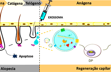
O ciclo capilar é um processo dinâmico e complexo que compreende a fase anágena de crescimento, catágena de regressão e telógena de queda.1,5
Sabe-se que as prostaglandinas influenciam o ciclo, uma vez que já foram identificados níveis altos de prostaglandina D2 e baixos de E2 por exemplo em pacientes com alopecia androgenética.6
Células mesenquimais progenitoras dos adipócitos tornam-se adipócitos maduros e passam a envolver o folículo durante a passagem da fase telógena para a anágena. Além disso são importantes para a proliferação celular após transplante capilar.1
A secreção de fatores de crescimento tipo FGF-5 produzidos pelas células da bainha externa ou o IGF-1 produzido pela célula da papila dérmica aumentam a proliferação folicular.1
Além disso, a ativação da via Wnt/ß-catenin também é fundamental para a manutenção do ciclo fisiológico e proliferação do folículo.5
Qualquer desregulação das vias de sinalização do ciclo capilar, seja por fatores ambientais, genéticos ou hormonais, pode levar a alopecia, uma condição que confere morbidade psicossocial ao paciente.4
Apesar das opções clínico-cirúrgicas atuais, o tratamento das alopecias ainda não é completamente satisfatório. Por isso, os EXM constituem uma possibilidade extremamente interessante.4,5
As células mesenquimais podem ser obtidas de vários tecidos e possuem alta capacidade de renovação e diferenciação.1 A interação entre células epiteliais e mesenquimais atua desde a diferenciação até a proliferação e migração das células foliculares por exemplo. Uma célula especializada de origem mesenquimal, a célula da papila dérmica (DP) regula a função, formação e crescimento do folículo, já que interfere na proliferação, migração e diferenciação das células da bainha externa (BE), cuja origem é epitelial.1,5
Os EXM de células DP podem promover a viabilidade das células DP, BE, células tronco foliculares, aumentam a expressão de fatores de crescimento e ativam cascatas que aceleram a passagem para a fase anágena e adiam a catágena. 2
Zhou e colaboradores conseguiram demonstrar o aumento da razão entre a fase anágena e catágena em ratos e estimular a proliferação, migração e expressão de B-catenina em células da BE utilizando EXM de cultura de células DP humanas saudáveis.7
Os exosomas derivados de cultura 3D de células da DP humanas aumentaram a porcentagem de células positivas para Ki67 em folículos pilosos cultivados e também a viabilidade de células da DP e células da BE, além de terem prolondado a fase anágena em ratos no estudo de Kwack e colaboradores.8
Yan e colaboradores identificaram mais de trinta RNA mensageiros expressos nos EXM de células da papila dérmica de cabra envolvidos na proliferação e diferenciação de células tronco folicular.9 Rajendran e colaboradores conseguiram demonstrar que EXM de células mesenquimais provocaram proliferação de células DP humanas e indução de VEGF e IGF-1.10 Também já foi identificada proliferação folicular in vivo a partir de EXM de células progenitoras de adipócitos evidenciados pela expressão de CD63, ALX1 e CD9 e levando também ao aumento da produção de VEGF e IGF-1.4
Nos estudos in vivo, os EXM são injetados e sua ação pode ser visualizada em documentação biográfica e mesmo em biópsias seriadas. Won e colaboradores obtiveram bons resultados em ratos tanto com a injeção quanto com a aplicação tópica de células tronco derivadas de adipócitos.11 Contudo, por mais promissor que seja o tema, mais estudos são necessários, uma vez que ainda existem muitas limitações em relação a identificação, isolamento e compreensão dos mecanismos de ação desses sinalizadores.
1 - Ha DH, Kim HK, Lee J, Kwon HH, Park GH, Yang SH, Jung JY, Choi H, Lee JH, Sung S, Yi YW, Cho BS. Mesenchymal Stem/Stromal Cell-Derived Exosomes for Immunomodulatory Therapeutics and Skin Regeneration. Cells. 2020 May 7;9(5):1157.
2 - Xiong M, Zhang Q, Hu W, Zhao C, Lv W, Yi Y, Wang Y, Tang H, Wu M, Wu Y. The novel mechanisms and applications of exosomes in dermatology and cutaneous medical aesthetics. Pharmacol Res. 2021 Apr;166:105490. doi: 10.1016/j.phrs.2021.105490. Epub 2021 Feb 12. PMID: 33582246.
3 - Wall D, Meah N, Fagan N, York K, Sinclair R. Advances in hair growth. Fac Rev. 2022 Jan 12;11:1. doi: 10.12703/r/11-1. PMID: 35156098; PMCID: PMC8808739
4 - Wu J, Yang Q, Wu S, Yuan R, Zhao X, Li Y, Wu W, Zhu N. Adipose-Derived Stem Cell Exosomes Promoted Hair Regeneration. Tissue Eng Regen Med. 2021 Aug;18(4):685-691.
5 - Hu S, Li Z, Lutz H, Huang K, Su T, Cores J, Dinh PC, Cheng K. Dermal exosomes containing miR-218-5p promote hair regeneration by regulating ß-catenin signaling. Sci Adv. 2020 Jul 24;6(30):eaba1685.
6 - Johnstone MA, Albert DM: Prostaglandin-Induced Hair Growth. Surv Ophthalmol. 2002; 47 Suppl 1: S185–S202.
7 - Zhou L, Wang H, Jing J, Yu L, Wu X, Lu Z. Regulation of hair follicle development by exosomes derived from dermal papilla cells. Biochem. Biophys. Res. Commun., 500 (2018), pp. 325-332
8 – Kwack MH, Seo CH, Gangadaran P, Ahn BC, Kim MK, Kim JC, Sung YK. Exosomes derived from human dermal papilla cells promote hair growth in cultured human hair follicles and augment the hair-inductive capacity of cultured dermal papilla spheres. Exp. Dermatol., 28 (2019), pp. 854-857
9 - Yan H, Gao Y, Ding Q, Liu J, Li Y, Jin M, Xu H, Ma S, Wang X, Zeng W, et al. Exosomal Micro RNAs Derived from Dermal Papilla Cells Mediate Hair Follicle Stem Cell Proliferation and Differentiation. Int. J. Biol. Sci. 2019, 15, 1368–1382.
10 - Rajendran, R.L.; Gangadaran, P.; Bak, S.S.; Oh, J.M.; Kalimuthu, S.; Lee, H.W.; Baek, S.H.; Zhu, L.; Sung, Y.K.; Jeong, S.Y.; et al. Extracellular vesicles derived from MSCs activates dermal papilla cell in vitro and promotes hair follicle conversion from telogen to anagen in mice. Sci. Rep. 2017, 7, 15560.
11 - Won CH, Yoo HG, Kwon OS, Sung MY, Kang YJ, Chung JH, Park BS, Sung JH, Kim WS, Kim KH. Hair growth promoting effects of adipose tissue-derived stem cells. J Dermatol Sci. 2010 Feb;57(2):134-7.
English version
Exosomes in hair restoration
Exosomes in hair restoration
Alessandro Alarcão
Gabriela Higino de Souza
Intercellular communication is the subject of research both for its diagnostic and therapeutic potential in the face of the body's physiological and pathological processes, and for a better elucidation of these processes. Synchronizing different cells at different times to carry out an in vivo process requires many coordinated signaling and response. Information transmission must occur between nearby cells and distant cells.
Around 1940, the scientific community faced the discovery of exosomes (EXM). However, it was not until the 2000s that these small vesicles (30 to 200 nm) surrounded by a phospholipid bilayer began to be deeply studied.1,2
Derived from intracellular multivesicular bodies, they are released by different cells in extracellular vesicles and may contain different substances.
These extracellular vesicles are classified according to nature, size, purity, action, quantity.¹ GTPases, annexins, CD63, CD81, CD9, heat shock proteins (HSP60, 70 and 90) among others were identified.²
Depending on the cell of origin, these structures may contain proteins, carbohydrates, lipids, nucleic acids, cytokines. Thus, they have varied sizes and functions, changing the biological response of the receptor cells, both stimulating and inhibiting. It is possible to modulate cellular actions such as proliferation, differentiation, migration, and death.2-4
Communication takes place with adjacent cells and at a distance, and as cells become senescent. Number and composition of these structures change, which also denotes their importance in aging.
The role of EXM is still not fully understood. EXM have already been linked to the pathophysiology of diseases such as cancer and inflammatory diseases. Thus, EXM can become therapeutic assets for regenerative and aesthetic medicine, from the prevention of scars and hyperchromia to rejuvenation and hair growth.1,2,5
The hair cycle is a dynamic and complex process comprising the anagen phase of growth, the catagen phase of regression and the telogen phase of loss.1,5
Prostaglandins influence the cycle, since elevated levels of prostaglandin D2 and low levels of E2 have been identified, for example, in patients with androgenetic alopecia.6
Mesenchymal progenitor cells of adipocytes become mature adipocytes and begin to surround the follicle during the transition from the telogen to anagen phase. In addition, they are important for cell proliferation after hair transplant.¹
Secretion of FGF-5-like growth factors produced by outer sheath cells or IGF-1 produced by dermal papilla cells enhance follicular proliferation.¹
Additionally, activation of the Wnt/ß-catenin pathway is also essential for maintaining the physiological cycle and follicle proliferation.5
Any deregulation of the hair cycle signaling, whether due to environmental, genetic, or hormonal factors, can lead to alopecia, a condition that provides psychosocial morbidity on the patient.4
Despite the current clinical-surgical options, the treatment of alopecia is still not completely satisfactory. Therefore, the EXM is an extremely interesting possibility.4,5
Mesenchymal cells can be obtained from various tissues and have a high capacity for renewal and differentiation.¹ The interaction between epithelial and mesenchymal cells acts from differentiation to proliferation and migration of follicular cells, for example. The dermal papilla (DP) cell is a specialized cell of mesenchymal origin and regulates the function, formation, and growth of the follicle, as it interferes with the proliferation, migration and differentiation of outer sheath (EB) cells, whose origin is epithelial.1,5
DP cell EXM can promote the viability of DP cells, BE, follicular stem cells, increase the expression of growth factors and activate cascades that accelerate the transition to the anagen phase and postpone the catagen phase. 2
Zhou et al could demonstrate an increase in the ratio between the anagen and catagen phase in mice and stimulate proliferation, migration and expression of B-catenin in EB cells using EXM from cultured healthy human DP cells.7
Exosomes derived from 3D culture of human PD cells increased the percentage of Ki67-positive cells in cultured hair follicles and the viability of PD cells and EB cells, in addition to prolonging the anagen phase in mice in the study by Kwack et al.8
Yan et al identified more than thirty messenger RNAs expressed in EXM of goat dermal papilla cells involved in follicular stem cell proliferation and differentiation.9 Rajendran et al could demonstrate that EXM from mesenchymal cells caused proliferation of human DP cells and induction of VEGF and IGF-1. Follicular proliferation has also been identified in vivo from EXM of adipocyte progenitor cells evidenced by the expression of CD63, ALX1 and CD9 and leading to increased production of VEGF and IGF-1.4
In in vivo studies, EXM are injected, and their action can be visualized in biographical documentation and even in serial biopsies. Won et al had reliable results in rats both with the injection and with the topical application of adipocyte-derived stem cells.11 However, as promising as the subject is, more studies are needed, since there are still many limitations in relation to identification, isolation and understanding of the mechanisms of action of these signals.
1 - Ha DH, Kim HK, Lee J, Kwon HH, Park GH, Yang SH, Jung JY, Choi H, Lee JH, Sung S, Yi YW, Cho BS. Mesenchymal Stem/Stromal Cell-Derived Exosomes for Immunomodulatory Therapeutics and Skin Regeneration. Cells. 2020 May 7;9(5):1157.
2 - Xiong M, Zhang Q, Hu W, Zhao C, Lv W, Yi Y, Wang Y, Tang H, Wu M, Wu Y. The novel mechanisms and applications of exosomes in dermatology and cutaneous medical aesthetics. Pharmacol Res. 2021 Apr;166:105490. doi: 10.1016/j.phrs.2021.105490. Epub 2021 Feb 12. PMID: 33582246.
3 - Wall D, Meah N, Fagan N, York K, Sinclair R. Advances in hair growth. Fac Rev. 2022 Jan 12;11:1. doi: 10.12703/r/11-1. PMID: 35156098; PMCID: PMC8808739
4 - Wu J, Yang Q, Wu S, Yuan R, Zhao X, Li Y, Wu W, Zhu N. Adipose-Derived Stem Cell Exosomes Promoted Hair Regeneration. Tissue Eng Regen Med. 2021 Aug;18(4):685-691.
5 - Hu S, Li Z, Lutz H, Huang K, Su T, Cores J, Dinh PC, Cheng K. Dermal exosomes containing miR-218-5p promote hair regeneration by regulating ß-catenin signaling. Sci Adv. 2020 Jul 24;6(30):eaba1685.
6 - Johnstone MA, Albert DM: Prostaglandin-Induced Hair Growth. Surv Ophthalmol. 2002; 47 Suppl 1: S185–S202.
7 - Zhou L, Wang H, Jing J, Yu L, Wu X, Lu Z. Regulation of hair follicle development by exosomes derived from dermal papilla cells. Biochem. Biophys. Res. Commun., 500 (2018), pp. 325-332
8 – Kwack MH, Seo CH, Gangadaran P, Ahn BC, Kim MK, Kim JC, Sung YK. Exosomes derived from human dermal papilla cells promote hair growth in cultured human hair follicles and augment the hair-inductive capacity of cultured dermal papilla spheres. Exp. Dermatol., 28 (2019), pp. 854-857
9 - Yan H, Gao Y, Ding Q, Liu J, Li Y, Jin M, Xu H, Ma S, Wang X, Zeng W, et al. Exosomal Micro RNAs Derived from Dermal Papilla Cells Mediate Hair Follicle Stem Cell Proliferation and Differentiation. Int. J. Biol. Sci. 2019, 15, 1368–1382.
10 - Rajendran, R.L.; Gangadaran, P.; Bak, S.S.; Oh, J.M.; Kalimuthu, S.; Lee, H.W.; Baek, S.H.; Zhu, L.; Sung, Y.K.; Jeong, S.Y.; et al. Extracellular vesicles derived from MSCs activates dermal papilla cell in vitro and promotes hair follicle conversion from telogen to anagen in mice. Sci. Rep. 2017, 7, 15560.
11 - Won CH, Yoo HG, Kwon OS, Sung MY, Kang YJ, Chung JH, Park BS, Sung JH, Kim WS, Kim KH. Hair growth promoting effects of adipose tissue-derived stem cells. J Dermatol Sci. 2010 Feb;57(2):134-7.