
Sandro Navarro Salanitri 1,2
Vinícius Cruz Prieto Silva 1,2
Anna Elisa Nóbrega de Souza 1,2
Christiane Steponavicius Sobral 1,2
1. Serviço de Cirurgia Plástica do Hospital Alemão Oswaldo Cruz Unidade Vergueiro
2. Sociedade Brasileira de Cirurgia Plástica, São Paulo, SP, Brasil.
Introdução
A cirurgia de implante capilar é considerada de baixo risco, com raras complicações e intercorrências. No entanto, quando qualquer ruído acontece, pode gerar uma enorme repercussão, angústia ao paciente e inclusive ao cirurgião; caso seja algo que ele não tenha visto antes, ou que não tenha sido documentado em artigos científicos.
Dr Sandro, mais uma vez, divide sua enorme experiência cirúrgica e deixa aqui registrado uma intercorrência atípica da cirurgia de implante capilar, com evolução favorável, porém prolongada. Registros como estes tranquilizarão cirurgiões que venham a experimentar uma situação semelhante.
Dra. Tatiana Tournieux
Resumo:
A cirurgia de transplante de cabelos pela técnica Folicular Unit Extraction (FUE), para muitos, é a cirurgia de eleição para o tratamento de alguns tipos de alopecia, principalmente a do tipo androgenética. Há poucos estudos sobre complicações decorrentes desta técnica cirúrgica na literatura médica, ao contrário do que ocorre na Técnica Follicular Unit Transplantation (FUT), provavelmente pelo fato daquela ser mais recente. Apresentamos o caso de um paciente que evoluiu com linfonodomegalia retroauricular bilateral pós transplante de cabelos por técnica FUE, que regrediu espontaneamente após 1 ano de cirurgia. Foram realizados exames laboratoriais e de imagem que não elucidaram a etiologia do processo.
Introdução:
Atualmente o transplante de cabelos (TC) pela técnica Folicular Unit Extraction (FUE) é a cirurgia mais realizada no mundo para o tratamento da alopecia androgenética (AA). A técnica Follicular Unit Transplantation (FUT), infelizmente, tem sido cada vez menos realizada, provavelmente em decorrência da maior procura do FUE pelos pacientes além da falta de conhecimento, prática e equipamentos necessários para a realização da técnica FUT por novos médicos e suas equipes). (1)
Como em todas as cirurgias, intercorrências e complicações podem ocorrer, e estas serão mais expressivas dependendo do número de procedimentos realizados pelo cirurgião, uma questão de estatística. A literatura médica sobre intercorrências e complicações da cirurgia de TC com a técnica FUE é pobre, ao contrário da técnica FUT, e tal assunto é de extrema importância para o diagnóstico prévio de possíveis complicações, o que amenizaria os possíveis prejuízos sofridos pelo paciente. (2-7)
O sistema imunológico humano tem a função de reconhecer e combater patógenos no organismos (desde um simples vírus até células neoplásicas malignas). Os gânglios linfáticos são responsáveis por barrar esses patógenos, sinalizando que algo de errado está ocorrendo, aumentando de tamanho acompanhado ou não de dor. Exames laboratoriais que evidenciem processo inflamatório ou infeccioso, sorologias, culturas, antibiograma, métodos de imagem, punções ou biópsias podem elucidar a causa do processo o órgão que acometido que está gerando o processo para o devido tratamento.(8, 9)
O organismo não interpreta uma cirurgia como benéfica, mas sim como uma agressão ao organismo. A tríplice reação de Lewis, dor, calor e rubor, é frequentemente observada em pós-operatório no sítio cirúrgico. Procedimentos cirúrgicos podem levar a linfonodomegalia, somente pelo processo inflamatório decorrente ao procedimento, que serão autolimitados.(10)
Tal relato de caso descreve um pós-operatório de TC por técnica FUE que evoluiu com linfonodomegalia retroauricular bilateral, que somente regrediram espontaneamente após 1 ano da cirurgia. O caso foi investigado com exames laboratoriais e de imagem não evidenciando a etiologia do processo, que na opinião do autor foi a agressão cirúrgica do TC. Não há relato na literatura de caso semelhante.
Descrição do Caso:
Paciente de raça branca, 34 anos, sem comorbidades ou alergias, negava cirurgias prévias, medicamentos ou vícios. Apresentava AA grau II, área doadora com cabelos espessos, escuros e ótima densidade pilosa. Decidiu realizar cirurgia de TC FUE para correção da AA. Foram solicitados exames laboratoriais (hemograma,coagulograma, glicemia, ureia e creatinina), além de eletrocardiograma, todos se mostraram normais, e o procedimento foi realizado em ambiente hospitalar.
Técnica cirúrgica empregada:
1. Documentação fotográfica
2. Depilação de couro cabeludo
3. Marcação de área doadora e receptora
4. Sedação com midazolam e fentanil aos cuidados do anestesiologista
5. Decúbito ventral
6. Assepsia e antissepsia
7. Anestesia local com xilocaína 20 ml + lidocaína 20 ml, ao redor da área doadora. Infiltração com xilocaína 20 ml + lidocaína 20 ml + adrenalina 1 ml + SF 0,9% 160 em área doadora.
8. Retira das unidades e famílias foliculares com Punch de 0,9 a 1 mm da área doadora demarcada, aproximadamente 1400 enxertos, manutenção destes em sf 0,9% resfriado a 4º graus.
9. Decúbito dorsal.
10. Anestesia local com xilocaína 20 ml + lidocaína 20 ml, ao redor da área receptora. Infiltração com xilocaína 20 ml + lidocaína 20 ml + adrenalina 1 ml + SF 0,9% 160 em área receptora.
11. Incisões prévias com lâmina de 0,5 mm em região receptora demarcada.
12. Enxertia das unidades e famílias foliculares.
13. Curativo.
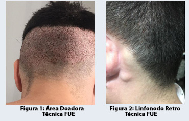
Paciente teve alta com analgésico se necessário e antibioticoterapia por 5 dias Retirado curativo no primeiro dia pós-operatório e retornos semanais. (figura 1). Nas primeiras semanas, o paciente reclamou de tumoração em região retroauricular bilateralmente. Sem queixas clínicas, apresentava linfonodomegalia de +/- 1 cm retroauricular bilateralmente. Restante do exame físico normal. Área receptora e doadora sem alterações, levantado hipótese diagnóstica de linfonodos reacionais decorrentes do procedimento cirúrgico. (figura 2)
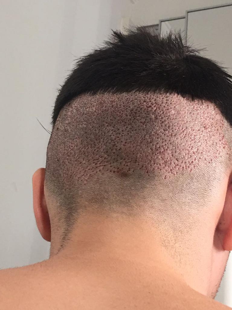
Figura 1: Área doadora técnica FUE
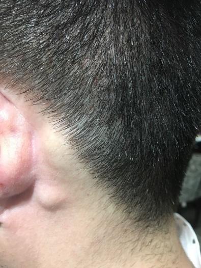
Figura 2: Linfonodo retro auricular
Com o passar do tempo, não houve regressão da linfonodomegalia. Paciente, por vontade própria, realizou ultrassonografia que confirmou a hipótese diagnóstica. Foram solicitados exames básicos de VHS, PCR e hemograma, que se mostraram sem alterações. Avaliação por infectologia descartou causa infecto-contagiosa.
A evolução da cirurgia ocorreu normalmente, com o eflúvio dos cabelos transplantados e crescimento ocorrendo devidamente após 3-6 meses da cirurgia. Durante o período, foi optado por observar os linfonodos comprometidos, que foram diminuindo progressivamente de tamanho até evoluírem totalmente após aproximadamente 1 ano após cirurgia. Paciente satisfeito com a cirurgia.
Discussão:
O sistema linfático é um sistema formado por vasos e órgãos linfoides por onde circula a linfa. É um sistema de drenagem auxiliar constituído pela medula óssea vermelha, timo, baço, linfonodos e vasos linfáticos que carregam a linfa drenada para o sistema venoso. (11)
Processos inflamatórios, infecciosos ou neoplásicos serão drenados para linfonodos do couro cabeludo. Topograficamente, os Ductos Linfáticos do couro cabeludo se dividem em:
- Os da região frontal: que terminam nos Linfonodos Auriculares Anteriores.
- Os da região temporo-parietal: que vão aos Linfonodos Parotídeos e Auriculares posteriores.
- Os da região occipital: que terminam parte nos Linfonodos occipitais, e parte nos Linfonodos cervicais profundos inferiores.
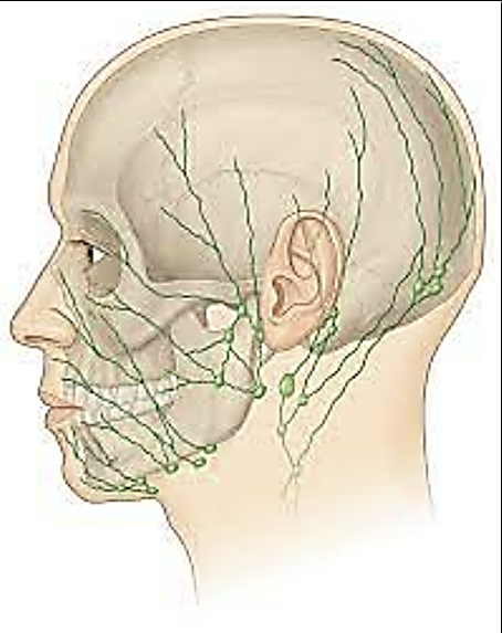
Figura 4: linfonodos do Cabeça.
Como já aventado, a literatura médica não faz citação específica do assunto em pauta. Em trabalho científico de 2009, sobre complicações e intercorrências em 533 cirurgias de transplante de cabelos, não é citado caso semelhante. No entanto, tal trabalho avalia somente a cirurgia FUT. Há possíveis motivos que valem ser discutidos: (7)
1. A cirurgia TC FUT é menos agressiva que a cirurgia TC FUE, visto que a área cruenta criada pelas inúmeras perfurações dos micro punchs é maior que as incisões necessárias para obtenção da fita de couro cabeludo, gerando menos processo inflamatório?
2. Para a realização da técnica FUE é obrigatório a depilação do couro cabeludo, retirando o cabelo ao contrário da FUT, que pode mascarar o processo.
3. A não valorização do processo tanto pelo paciente quanto pelo médico, visto que tal linfonodomegalia é de resolução espontânea e rápida, diferente do ocorrido neste caso (que foi duradoura), o que levou a investigação clínica.
Apesar de, na maioria das vezes, a etiopatogenia do processo ser a agressão cirúrgica, não podemos descartar outros motivos para tumorações no área doadora do TC e sua vizinhança.
Devemos lembrar que outras possíveis tumorações podem aparecer durante o pós-operatório de transplante de cabelos: desde pequenos cistos decorrentes de cabelos encravados, até fístulas artério-venosas descritas em literatura. (12, 13) - verifique o artigo “Fístula arteriovenosa pós-transplante de cabelos: Relatos de casos”, publicado em agosto de 2021, na edição 22 de O Folículo. Portanto, para o diagnóstico no caso de dúvida, o melhor exame (menos agressivo) é o USG doppler que pode evidenciar fluxo dentro da tumoração, elucidando o diagnóstico e norteando a terapêutica.
No caso presente, a involução dos linfonodos, apesar de tardia, ocorreu de forma espontaneamente. A avaliação de outros profissionais, sejam infectologistas ou cirurgiões de cabeça e pescoço, pode se fazer necessária, uma vez que outras patologias também podem levar a adenomegalia, comprometendo a saúde do paciente. (9)
Conclusão:
Linfonodomegalia pode ocorrer após TC FUE em decorrência a agressão cirúrgica. Na maioria das vezes, são autolimitadas e se resolvem em curto espaço de tempo. Em casos mais arrastados, devemos investigar outras possíveis causas de tumoração em couro cabeludo, desde as decorrentes do processo cirúrgico propriamente dito, até as causas infecciosas/neoplásicas. Estudos sobre complicações de TC FUE devem ser realizados.
Bibliografia:
1. Lam SM. Hair Transplant 360-Volume 3: Advances, Techniques, Business Development & Global Perspectives: JP Medical Ltd; 2014.
2. Kerure AS, Patwardhan N. Complications in Hair Transplantation. J Cutan Aesthet Surg. 2018;11(4):182-9.
3. Lam SM. Complications in hair restoration. Facial Plast Surg Clin North Am. 2013;21(4):675-80.
4. Loganathan E, Sarvajnamurthy S, Gorur D, Suresh DH, Siddaraju MN, Narasimhan RT. Complications of hair restoration surgery: a retrospective analysis. Int J Trichology. 2014;6(4):168-72.
5. Nadimi S. Complications with Hair Transplantation. Facial Plast Surg Clin North Am. 2020;28(2):225-35.
6. Perez-Meza D, Niedbalski R. Complications in hair restoration surgery. Oral Maxillofac Surg Clin North Am. 2009;21(1):119-48, vii.
7. Salanitri S, Goncalves AJ, Helene A, Jr., Lopes FH. Surgical complications in hair transplantation: a series of 533 procedures. Aesthet Surg J. 2009;29(1):72-6.
8. Chammas MC, Lundberg JS, Juliano AG, Saito OdC, Marcelino ASZ, Cerri GG. Linfonodos cervicais: um dilema para o ultra-sonografista. Radiologia Brasileira. 2004;37:357-64.
9. Crespo A, Meléndez A, Montovani J, Cavinato J, Odone Filho V. Adenopatias Cervicais. IV Manual de otorrinolaringologia pediátrica da IAPO Guarulhos: Lis gráfica & Editora. 2006:93-101.
10. Balbino CA, Pereira LM, Curi RJRBdCF. Mecanismos envolvidos na cicatrização: uma revisão. 2005;41(1):27-51.
11. Gardner E, Gray DJ, O'Rahilly R, Benevento R. Anatomia: estudo regional do corpo humano. Anatomia: estudo regional do corpo humano: Guanabara Koogan; 1978.
12. Bernstein J, Podnos S, Leavitt M. Arteriovenous fistula following hair transplantation. Dermatologic surgery. 2011;37(6):873-5.
13. LEÃO¹ CEG, Goulart BCT, Fonseca TG, RASSI SP. Pseudoaneurisma da artéria occipital em área doadora de cirurgia de calvície. Rev Soc Bras Cir Plást. 2005;20(1):59-62.
Edição 26, artigo 3.
English version
Case description of retroauricular lymphadenomegaly after hair transplant surgery using the Follicular Unit Extraction (FUE) technique
Sandro Navarro Salanitri 1,2
Vinícius Cruz Prieto Silva 1,2
Anna Elisa Nóbrega de Souza 1,2
Christiane Steponavicius Sobral 1,2
1. Serviço de Cirurgia Plástica do Hospital Alemão Oswaldo Cruz Unidade Vergueiro
2. Sociedade Brasileira de Cirurgia Plástica, São Paulo, SP, Brasil.
Introduction
Hair implant surgery is considered minimal risk, with rare complications and intercurrences. However, when any noise occurs, it can generate enormous repercussions, anguish for the patient and even the surgeon; if it is something he has not seen before, or something that has not been documented in scientific papers.
Once again, Dr Sandro shares his enormous surgical experience and records here an atypical intercurrence of hair implant surgery, with favorable but prolonged evolution. Records like these will reassure surgeons who might experience ananalogous situation.
Dr. Tatiana Tournieux
Abstract:
For many people, hair transplant surgery using the Follicular Unit Extraction (FUE) technique is the surgery of choice for the treatment of some types of alopecia, especially the androgenetic type. There are few studies on complications resulting from this surgical technique in the medical literature, contrary to what happens in the Follicular Unit Transplantation (FUT) technique, probably because it is a more recent technique. We present the case of a patient who evolved with bilateral retroauricular lymph node enlargement after hair transplantation using the FUE technique, which spontaneously regressed after one year of surgery. Laboratory tests and imaging tests were performed, and the etiology of the process was not clarified.
Summary:
Hair transplant surgery using the Follicular Unit Extraction (FUE) technique, for many, is a main surgery of choice for the treatment of some types of alopecia, especially androgenetic alopecia. There are few studies about complications of FUT in the medical literature, unlike that occurs in the Follicular Unit Transplantation Technique, probably because it is the latest technique. We present a case of a patient that evolved with bilateral retroauricular lymphonodemegaly after CT using FUE technique, which resolved spontaneously after 1 year of surgery. Laboratory and imaging tests were performed, which did not elucidate the ethology of the process.
Introduction:
Currently, hair transplant (HT) using the Follicular Unit Extraction (FUE) technique is the most performed surgery in the world for the treatment of androgenetic alopecia (AA). Unfortunately, the Follicular Unit Transplantation (FUT) technique has been less and less performed, due to the greater demand for FUE by patients in addition to the lack of knowledge, practice, and equipment necessary for the performance of the FUT technique by new doctors and their teams). (1)
As in all surgeries, intercurrences and complications can occur, and these will be more expressive depending on the number of procedures performed by the surgeon. It is a matter of statistics. Medical literature on complications and complications of hair transplant surgery with the FUE technique is poor, unlike the FUT technique, and this subject is extremely important for the prior diagnosis of complications, which would mitigate the possible harm suffered by the patient. (2-7)
The human immune system has the function of recognizing and fighting pathogens in the organism (from a simple virus to malignant neoplastic cells). Lymph nodes are responsible for blocking these pathogens, signaling that something is wrong, increasing in size with or without pain. Laboratory tests that show an inflammatory or infectious process, serologies, cultures, antibiogram, imaging methods, punctures or biopsies can elucidate the cause of the process, and the affected organ that is generating the process for proper treatment.(8, 9) The organism does not interpret a surgery as beneficial, but as an aggression.
The triple Lewis reaction (pain, heat, and redness) is frequently observed postoperatively at the surgical site. Surgical procedures can lead to lymph node enlargement, just because of the inflammatory process resulting from the procedure, which will be self-limited.(10)
This case report describes a postoperative period of hair transplant using the FUE technique that evolved with bilateral retroauricular lymphadenopathy, which only spontaneously regressed one year after surgery. The case was investigated with laboratory and imaging tests not showing the etiology of the process. In the author's opinion, it was the surgical aggression of the hair transplant. There is no report of a similar case in the literature.
Case description:
Caucasian patient, 34 years old, without comorbidities or allergies, denied previous surgeries, medication, or addictions. She had grade II AA, a donor area with thick, dark hair and excellent hair density. Decided to perform hair transplant FUE surgery for AA correction. Laboratory tests were requested (blood count, coagulogram, blood glucose, urea, and creatinine), in addition to electrocardiogram, all of which were normal, and the procedure was performed in a hospital.
Surgical technique used:
1. Photographic documentation
2. Scalp hair removal
3. Donor and recipient area marking
4. Sedation with midazolam and fentanyl under the care of the anesthesiologist
5. Prone position
6. Asepsis and antisepsis
7. Local anesthesia with 20 ml xylocaine + 20 ml lidocaine around the donor area. Infiltration with 20 ml xylocaine + 20 ml lidocaine + 1 ml adrenaline + 0.9% SF 160 in the donor area.
8. Removal of the follicular units and follicular families with a 0.9 to 1 mm Punch from the demarcated donor area, approximately 1400 grafts, maintenance of these in sf 0.9% cooled to 4º degrees.
9. Supine position.
10. Local anesthesia with xylocaine 20 ml + lidocaine 20 ml, around the receiving area. Infiltration with 20 ml xylocaine + 20 ml lidocaine + 1 ml adrenaline + 0.9% SF 160 in the receiving area.
11. Previous incisions were made with a 0.5 mm blade in the demarcated recipient region.
12. Grafting of follicular units and families.
13. Dressing.
Patient was discharged with analgesics if necessary and antibiotic therapy for 5 days. Dressing removed on the first postoperative day and weekly returns. (Figure 1). In the first weeks, the patient complained of a tumor in the retroauricular region bilaterally. Without clinical complaints, he had lymph node enlargement of +/- 1 cm bilaterally retroauricular. Remainder of normal physical examination. Recipient and donor area without alterations, raising the diagnostic hypothesis of reactive lymph nodes resulting from the surgical procedure. (Figure 2)
Figure 1: Donor area - FUE technique
Figure 2: Retro auricular lymph node
Over time, there was no regression of lymph node enlargement. The patient, voluntarily, underwent an ultrasound that confirmed the diagnostic hypothesis. Basic tests of ESR, CRP and blood count were requested, which showed no alterations. Evaluation by infectiology ruled out an infectious-contagious cause.
The evolution of the surgery occurred normally, with the effluvium of the transplanted hair and growth occurring properly after 3-6 months of the surgery. During the period, it was decided to observe the compromised lymph nodes, which progressively decreased in size until they fully evolved approximately one year after surgery. Patient satisfied with the surgery.
Discussion:
The lymphatic system is formed by vessels and lymphoid organs through which the lymph circulates. It is an auxiliary drainage system consisting of the red bone marrow, thymus, spleen, lymph nodes and lymphatic vessels that carry drained lymph to the venous system. (11)
Inflammatory, infectious, or neoplastic processes will drain into scalp lymph nodes. Topographically, the lymphatic ducts of the scalp are divided into:
- Those in the frontal region end in the anterior auricular lymph nodes.
- Those of the parietal and temporal region go to the parotid and posterior auricular lymph nodes.
- Those of the occipital region partly terminate in the occipital lymph nodes, and partly in the inferior deep cervical lymph nodes.
Figure 4: Head lymph nodes.
As already suggested, the medical literature does not specifically mention the subject in question. In a scientific paper from 2009, on complications and intercurrences in 533 hair transplant surgeries, no similar case is mentioned. However, this paper only evaluates the FUT surgery. There are reasons worth discussing: (7)
1. Is the FUT surgery less aggressive than the FUE surgery, since the bloody area created by the numerous perforations of the micro punches is larger than the incisions necessary to obtain the scalp tape, generating less inflammation?
2. To perform the FUE technique, it is mandatory to remove hair from the scalp, unlike FUT, which can mask the process.
3. The non-appreciation of the process by both the patient and the doctor, since such lymphadenopathy has a spontaneous and rapid resolution, unlike what happened in this case (which was long-lasting), which led to clinical investigation.
Although, in most cases, the etiopathogenesis of the process is surgical aggression, we cannot rule out other reasons for tumors in the hair transplant donor area and its vicinity. We must remember that other tumors may appear during the hair transplant postoperative period: from small cysts resulting from ingrown hairs to arteriovenous fistulas described in the literature. (12, 13) - check article “Arteriovenous fistula after hair transplantation: Case reports”, published on August, 2021, issue 22, O Folículo. Therefore, for the diagnosis in case of doubt, the best exam (less aggressive) is the USG doppler that can show flow inside the tumor, elucidating the diagnosis and guiding the therapy.
In the present case, the involution of the lymph nodes, occurred spontaneously, although late. The evaluation of other professionals, whether infectologists or head and neck surgeons, may be necessary, since other pathologies can also lead to adenomegaly, compromising the patient's health. (9)
Conclusion:
Lymph node enlargement may occur after FUE technique due to surgical aggression. Most of the time, they are self-limited and resolve in a brief period. In longer-lasting cases, we should investigate other probable causes of a tumor on the scalp, from those resulting from the surgical process itself, to nfectious/neoplastic causes. Studies on complications of CT FUE should be performed.
Bibliografia:
1. Lam SM. Hair Transplant 360-Volume 3: Advances, Techniques, Business Development & Global Perspectives: JP Medical Ltd; 2014.
2. Kerure AS, Patwardhan N. Complications in Hair Transplantation. J Cutan Aesthet Surg. 2018;11(4):182-9.
3. Lam SM. Complications in hair restoration. Facial Plast Surg Clin North Am. 2013;21(4):675-80.
4. Loganathan E, Sarvajnamurthy S, Gorur D, Suresh DH, Siddaraju MN, Narasimhan RT. Complications of hair restoration surgery: a retrospective analysis. Int J Trichology. 2014;6(4):168-72.
5. Nadimi S. Complications with Hair Transplantation. Facial Plast Surg Clin North Am. 2020;28(2):225-35.
6. Perez-Meza D, Niedbalski R. Complications in hair restoration surgery. Oral Maxillofac Surg Clin North Am. 2009;21(1):119-48, vii.
7. Salanitri S, Goncalves AJ, Helene A, Jr., Lopes FH. Surgical complications in hair transplantation: a series of 533 procedures. Aesthet Surg J. 2009;29(1):72-6.
8. Chammas MC, Lundberg JS, Juliano AG, Saito OdC, Marcelino ASZ, Cerri GG. Linfonodos cervicais: um dilema para o ultra-sonografista. Radiologia Brasileira. 2004;37:357-64.
9. Crespo A, Meléndez A, Montovani J, Cavinato J, Odone Filho V. Adenopatias Cervicais. IV Manual de otorrinolaringologia pediátrica da IAPO Guarulhos: Lis gráfica & Editora. 2006:93-101.
10. Balbino CA, Pereira LM, Curi RJRBdCF. Mecanismos envolvidos na cicatrização: uma revisão. 2005;41(1):27-51.
11. Gardner E, Gray DJ, O'Rahilly R, Benevento R. Anatomia: estudo regional do corpo humano. Anatomia: estudo regional do corpo humano: Guanabara Koogan; 1978.
12. Bernstein J, Podnos S, Leavitt M. Arteriovenous fistula following hair transplantation. Dermatologic surgery. 2011;37(6):873-5.
13. LEÃO¹ CEG, Goulart BCT, Fonseca TG, RASSI SP. Pseudoaneurisma da artéria occipital em área doadora de cirurgia de calvície. Rev Soc Bras Cir Plást. 2005;20(1):59-62.
Issue 26, artigo 3.